Heat pain in the spinal cord
Chronic pain affects millions, yet we still don't fully understand how pain signals travel through the spinal cord. This project uses ultra high field MRI to visualize those pathways with unprecedented detail.

The spinal cord: the body's overlooked pain processor
Most people are familiar with brain scans, but far fewer know that we can also measure activity in the spinal cord, the highway that carries every pain signal from the body to the brain. For pain research, this region is crucial: when you touch something painfully hot, the signal doesn't just travel through the spinal cord, it's actively shaped and modulated there.
The Challenge
The spinal cord is tiny, surrounded by bone, and constantly moving with every breath and heartbeat.
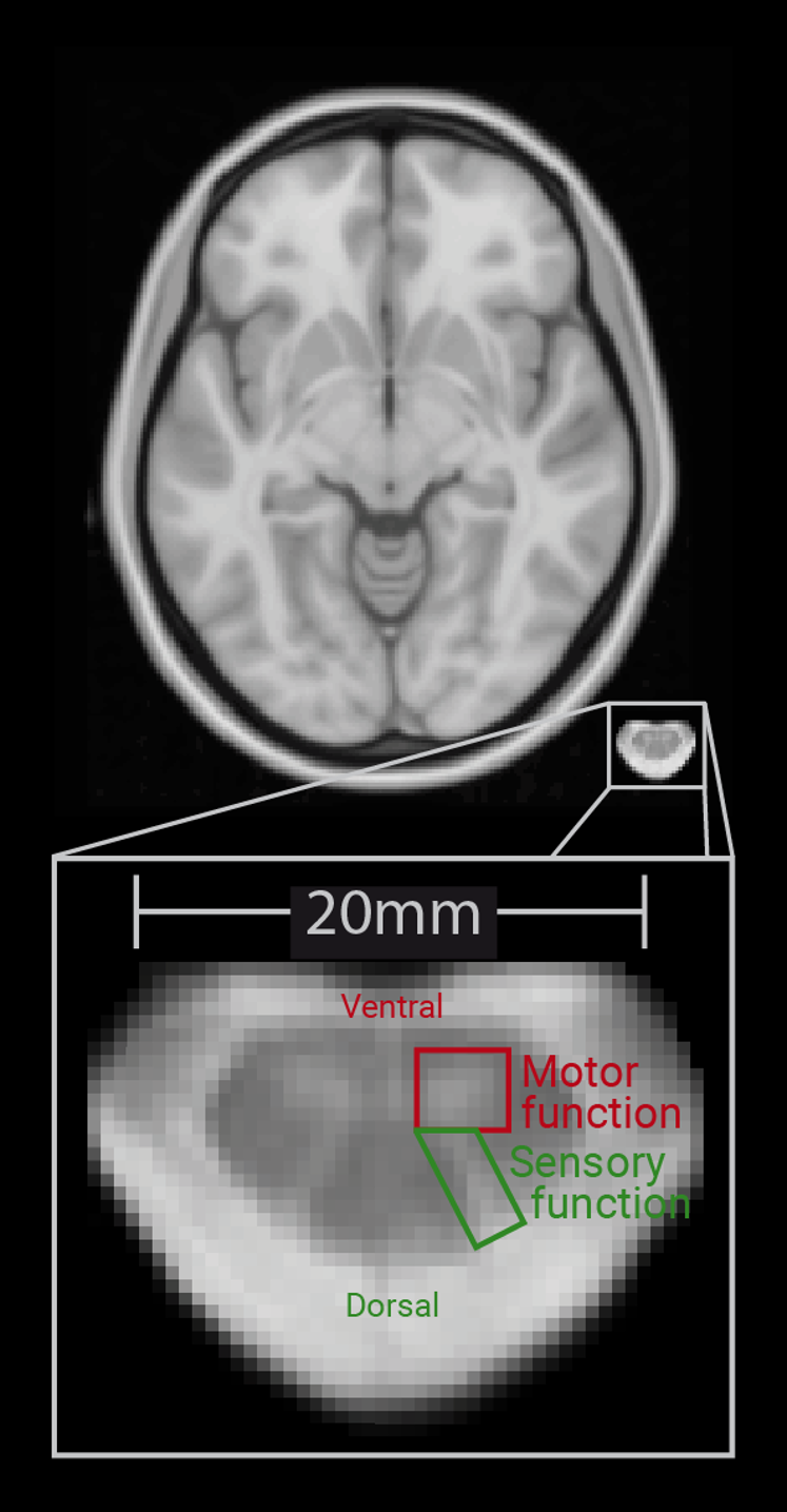
The current level of detail that standard MRI scanners with 3 Tesla can provide is barely enough to even disentangle motor and sensory regions within the spinal cord.
To study spinal cord pain processing, we need imaging technology that is both ultra precise and powerful enough to distinguish details only millimeters apart.
But at the start of the project task-based 7 Tesla fMRI of the spinal cord has not been done successfully yet.
Approach
- We worked with a 7 Tesla MRI scanner, which allows for much finer detail than standard clinical scanners.
- Because no suitable spinal cord coil exists at this field strength, we used a custom-built coil designed specifically for this project.
- Participants received precisely controlled heat stimuli on the skin to trigger real pain signals.
- We applied advanced imaging and preprocessing techniques to capture clean spinal cord activity despite artifacts, breathing and movement.
- We created a high resolution spinal cord atlas to pinpoint exactly where these responses occurred.
- Finally, we measured how the spinal cord reacts to incoming pain signals in real time.
What I contributed
- I designed and programmed the entire experimental protocol, including thermode control and its integration with PsychoPy.
- I conducted a large pre-study with 62 participants and a main study with 41 measurements — more than 100 spinal cord MRI sessions in total.
- I preprocessed all MRI data using FSL and the Spinal Cord Toolbox, building reproducible bash workflows for motion correction and denoising.
- I developed analysis pipelines and visualized the results in Python.
- I wrote the manuscript, including the literature review and interpretation of the findings.
- I created publication-ready figures in Adobe Illustrator.
- I documented the full workflow with reproducible code on GitHub.
Research & Analysis Stack
What we found
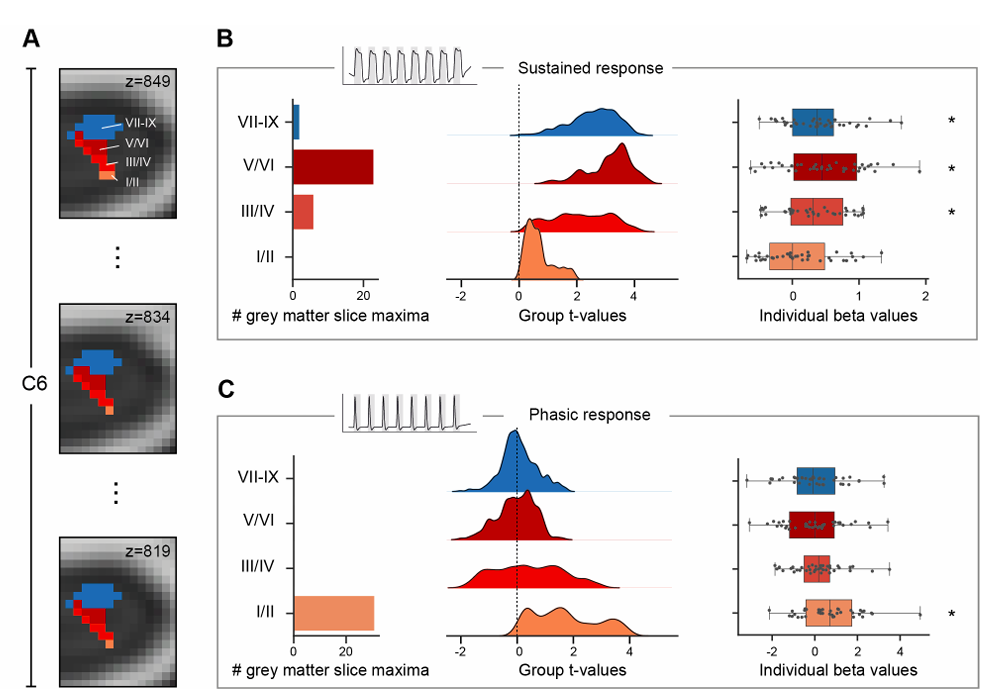
We were able to measure clear pain related activity in the dorsal horn, the part of the spinal cord where incoming nociceptive signals are processed. These responses showed a consistent spatial pattern and were strong enough to detect not only across the group, but even in individual participants.
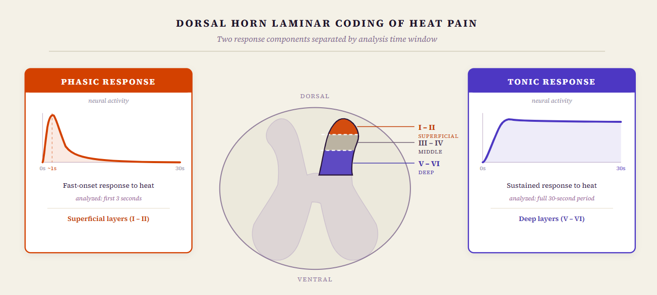
To understand how the spinal cord handles different aspects of heat pain, we tested two components of the response by analyzing different time windows of the same stimulus: a fast, phasic response during the first three seconds, and a slower, tonic response during the full 30 second heat period.
By separating these two components, we could see where in the dorsal horn each type of signal was strongest. The phasic response — the quick reaction to the onset of heat — was most prominent in the superficial layers of the dorsal horn. The tonic response — the sustained signal during ongoing heat — appeared more strongly in the deep layers.
This layer specific pattern shows that the spinal cord organizes incoming pain information by depth: fast, sharp signals at the surface; slower, ongoing signals deeper inside. Demonstrating this separation clearly with human spinal fMRI provides a new way to study early pain processing.